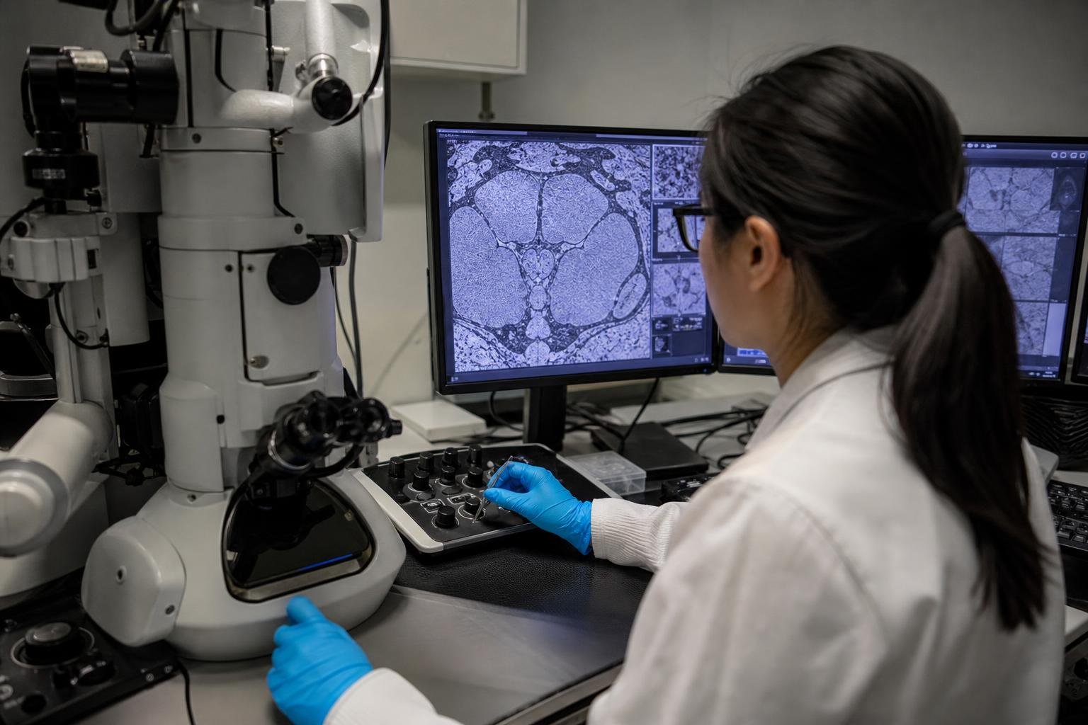
Google Research and the Howard Hughes Medical Institute (HHMI) have released a complete three-dimensional map of the adult male fruit fly brain and central nervous system, covering more than 166,000 neurons. The searchable reconstruction was built from millions of electron-microscopy image slices with AI-assisted methods, turning raw tissue imagery into a shared reference for studying neural circuits.[1]
The significance is not simply the scale of the dataset. With a previously mapped female fruit fly brain available for comparison, researchers now have whole-brain references for both sexes of the same model organism. That gives neuroscience a more direct way to investigate which differences in neural wiring may be linked to sex-specific behavior, rather than inferring those differences from partial brain regions or isolated cell types.[1]
By the numbers
- More than 166,000: neurons reconstructed in the adult male fruit fly brain and central nervous system.
- Millions: imaging slices processed to create the reconstruction.
- Two: whole-brain reference maps available across sex: male and previously mapped female fruit flies.
- One: searchable three-dimensional connectome intended as a scientific reference resource.
A matched reference for studying behavior
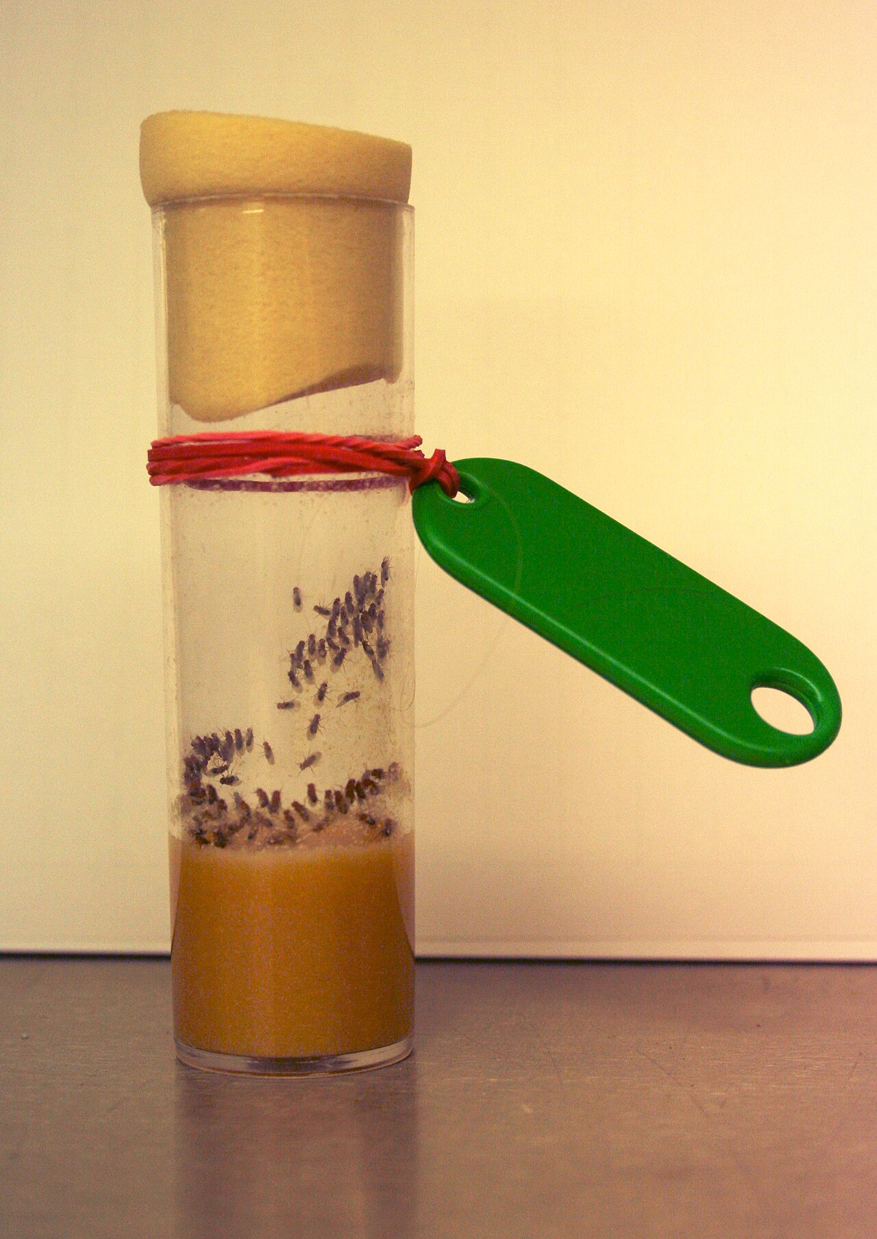
Fruit flies, Drosophila melanogaster, have long been a central model organism in biology. They are experimentally tractable, share many fundamental cellular and genetic processes with other animals, and exhibit measurable behaviors involving courtship, aggression, feeding, sleep, navigation and learning. Their nervous systems are far smaller than mammalian brains, but remain complex enough to make full circuit-level analysis technically difficult.
A connectome is a map of neural components and their connections. In practice, producing one requires more than imaging a brain. Researchers must identify the boundaries of individual neurons across a vast stack of extremely detailed tissue images, trace their branches through the volume, and organize the resulting cells and connections into data other scientists can query.
The male map changes the scientific value of the existing female reference because it creates a matched comparison rather than an isolated catalog. Researchers can now ask whether a circuit associated with a behavior is structurally conserved across sexes, differs in cell number or connectivity, or contains specialized pathways. Such comparisons are especially relevant for behaviors that show clear sex dependence, including reproductive and social behaviors.
Importantly, a structural difference is not by itself proof of a behavioral cause. Circuit function also depends on neural activity, chemical signaling, developmental history and environmental conditions. The new resource narrows the search space: it identifies candidate wiring differences that can be tested with genetics, imaging and behavioral experiments.
AI made the reconstruction tractable
The central engineering achievement is the use of AI-assisted reconstruction to handle an imaging problem that would otherwise be overwhelming. Electron microscopy can capture the fine structures needed to distinguish neighboring neurons, but it produces enormous numbers of two-dimensional slices. To build a usable three-dimensional brain map, each cellular process must be followed correctly from slice to slice.
Machine-learning systems can segment an electron-microscopy volume by predicting which pixels or voxels belong to the same underlying structure. Those predictions make it possible to assemble candidate neurons at a scale that manual tracing alone cannot support. The resulting workflow is not a matter of pressing a button and accepting a finished map. Automated segmentation can make merge errors, in which separate neurons are joined, or split errors, in which one neuron is broken into multiple fragments. Quality control, proofreading and biological validation remain necessary parts of producing a reliable reference.
Google Research describes the release as an AI-assisted effort that converts millions of imaging slices into a navigable, searchable three-dimensional connectome.[1] That is the practical role of AI in this project: not a model claiming to explain cognition, but a high-throughput reconstruction tool that makes foundational biological data usable by a broader research community.
This distinction matters. AI is increasingly valuable in scientific workflows where the bottleneck is interpreting large, repetitive and high-resolution datasets. Connectomics is an especially clear example because the raw data are visually rich but too voluminous for conventional manual annotation. The output is durable infrastructure: a reference dataset that can support many research questions beyond the original reconstruction project.
Key players and the infrastructure model
Google Research and HHMI are the principal organizations behind the announcement.[1] HHMI’s research ecosystem has played a prominent role in advanced biological imaging and neuroscience, while Google contributes machine-learning and large-scale data-processing expertise. Their collaboration reflects a broader pattern in modern life science: progress increasingly depends on teams that combine microscopy, neuroanatomy, software engineering, machine learning, data systems and expert annotation.
The public value of the work depends heavily on accessibility. A connectome becomes far more useful when researchers can search for cells, inspect morphology, compare regions and connect anatomical observations to experimental data. The announcement frames the male map as a resource for the community rather than a result intended only for a single laboratory.[1]
That shared-infrastructure approach can change how smaller neuroscience groups operate. Instead of undertaking years of reconstruction work before testing a hypothesis, a laboratory may begin with a candidate circuit identified in the reference map, then use targeted experiments to test what that circuit does. The map cannot replace experiments, but it can reduce the cost and time required to formulate more precise ones.
What the release means for AI and neuroscience
For the AI industry, the project is evidence that the most consequential uses of machine learning may be embedded in specialized scientific pipelines rather than presented as standalone consumer products. The relevant measures are not only model benchmarks. They include reconstruction accuracy, error-detection workflows, throughput, interoperability and whether outside researchers can use the resulting data product.
For microscopy and research-software providers, complete connectomes also raise demand for tools that can manage massive volumetric datasets, support collaborative proofreading, link anatomy to gene-expression and activity data, and make complex biological structures intuitive to explore. The commercial effect is likely to be indirect and gradual: publicly produced reference maps can create standards and technical expectations that influence academic instrumentation, cloud computing and scientific visualization markets.
The broader research opportunity is comparative connectomics. Once maps from different sexes, developmental stages, genetic backgrounds or species can be aligned, scientists can move from asking what a single nervous system contains to asking how circuits vary. The male and female fruit fly references are a meaningful early foundation for that approach because the comparison is whole-brain in scope.
Limits, concerns and the next technical hurdles
Completeness should not be confused with finality. A connectome is a detailed structural snapshot of a particular specimen and preparation. Biological variation between individual flies remains important, and a single reference does not establish the full range of normal wiring patterns. Comparing male and female maps also requires careful alignment of corresponding cell types and regions; apparent differences can arise from biological variation, annotation choices or reconstruction uncertainty.
There is also a risk of overinterpreting anatomy. Neural connections reveal possible information pathways, not necessarily the strength, timing or context of signaling through those pathways. Functional measurements remain essential to establish whether a structural difference changes behavior. Researchers will need to combine the map with optogenetics, calcium imaging, genetics and behavioral assays to turn candidate differences into causal findings.
Data scale remains a practical concern. Large electron-microscopy datasets demand substantial storage, compute and specialized visualization tools. Open access is most valuable when it is accompanied by documentation, stable identifiers, interoperable formats and mechanisms for reporting and correcting errors. AI can accelerate reconstruction, but it also makes provenance and quality assessment more important: users need to know how a neuronal segment was generated, reviewed and revised.
The next milestone is likely to be less about a larger single map than about making comparisons routine. Researchers will look for tools that match cells across the male and female references, quantify wiring differences, flag uncertain segments and link anatomy to experiments. If that layer of analysis becomes reliable, the connectomes can evolve from static atlases into active platforms for hypothesis generation.
Editor’s Take
I see the paired male-and-female reference as the real product here. A 166,000-neuron reconstruction is impressive, but the practical payoff comes when a researcher can ask a comparative question at whole-brain scale and get a tractable list of circuits to test. That is a far better use of AI than treating a generative model as a substitute for experimental biology.
The next thing to watch is whether the data and analysis tools make cross-sex comparison genuinely routine for labs that did not build the map. If they do, this becomes infrastructure with compounding value. The hype would outrun the facts if anyone suggests the map explains behavior on its own; anatomy provides hypotheses, while causal experiments still supply answers.